[ad_1]

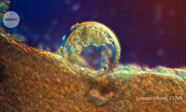
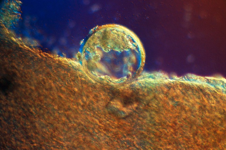
A human embryo at just three days old, implanted in the uterus. The cells need a store of proteins to kick start their development.Credit: Lennart Nilsson, TT/Science Photo Library
For almost 60 years, scientists have puzzled over the purpose of bundles of fibres that were found floating in mammalian egg cells. A study1 published today in Cell finds that these fibres, known as cytoplasmic lattices, are storage sites for many proteins that are essential for the development of the early embryo. The discovery could explain why people whose eggs lack the fibres entirely are infertile.
“A lot of these proteins have really important functions for the early mammalian embryo,” says Melina Schuh, a biochemist at the Max Planck Institute for Multidisciplinary Sciences in Göttingen, Germany, and an author of the paper. She says she has not seen a protein-storage system like it in mammalian cells before. “It is a very different, unique mechanism.”
Scientists first imaged cytoplasmic lattices in the 1960s using electron microscopy and thought they were flat sheets of fibres stacked around the nucleus. But using cryo-electron tomography, a microscopy technique in which the sample is frozen and scanned, Ida Jentoft, another co-author and a biochemistry graduate student at the Max Planck Institute for Multidisciplinary Sciences, and her colleagues saw that the lattices were not sheets after all, and in more of a bundle than a lattice.
They imaged mouse egg cells, known as oocytes, at a resolution of 30 ångströms — about the length of one twist of a DNA molecule — in three dimensions. Each fibre of the cytoplasmic lattice was, on average 7,000 ångströms long and 1,000 ångströms wide, and was composed of elliptical filaments stacked together in a staggered manner. The staggered stacking increases the surface area for protein storage, says Jentoft. Each fibre had between 5 and 40 filaments. “There’s not one recipe,” she says.
Scott Coonrod, a biologist at Cornell University’s Baker Institute for Animal Health in Ithaca, New York, says the paper reveals the molecular underpinnings of cytoplasmic lattices and shows how essential they are in the early embryo. “Importantly, this study provides us with the first high-resolution structural analysis of these filaments,” he says.
Fertility clues
The findings provide a new insight into embryo development. Mature oocytes contain material, derived from the mother, that they use to divide and grow after fertilization. An oocyte might not be fertilized immediately on maturation, so the material needs to be stored. That poses a problem because all cells, including oocytes, have processes that break down and recycle unused proteins. But the oocyte solves this problem by attaching proteins to the cytoplasmic lattices, where they seem to be exempt from the recycling processes, Schuh suggests.
To identify what the cytoplasmic lattice is made of, Jentoft and her colleagues used expansion microscopy, a new technique that allows large and complex cells, such as oocytes, to be physically magnified and imaged in fine detail using conventional fluorescence microscopes. They found that the filaments are probably composed of proteins called PADI6 and SCMC, among others.
Disabling the genes that produce PADI6 and SCMC disrupted the cytoplasmic lattices and protein storage, resulting in nonviable embryos. “It was a revelation,” Schuh says.
Hiroyuki Sasaki, a geneticist at Kyushu University in Fukuoka, Japan, says the study shows that PADI6 and SCMC proteins are distributed throughout the cell. Scientists had previously thought the proteins were present only closer to the outer membrane.
The team confirmed that the findings also apply to human oocytes, providing an avenue for a better understanding of some forms of infertility. Fertilized eggs in people who have mutations in these genes usually do not progress past the first division. In vitro fertilization will not help, Jentoft says. “Knowing that IVF is not going to be an option if you have those mutations is very powerful information to have, because going through the cycles as a couple is extremely emotionally draining and expensive.”
Source link


